Detecting COVID-19 from CT Scans Using Deep Learning: A Comprehensive Review
Introduction
The COVID-19 pandemic has underscored the vital importance of rapid and accurate diagnostic tools to contain the spread of the virus and guide clinical management. While reverse transcription polymerase chain reaction (RT-PCR) remains the gold standard for confirming SARS-CoV-2 infection, it has several limitations including long turnaround times, high false negative rates, and limited accessibility in some regions [1].
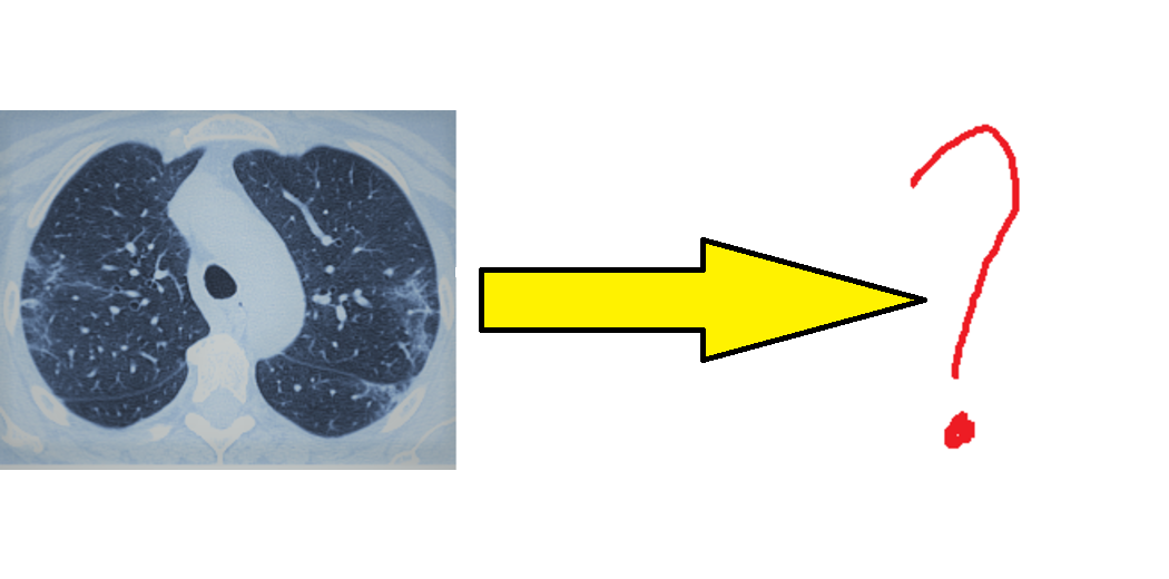
Chest computed tomography (CT) has emerged as a complementary tool for diagnosing COVID-19, especially in early stages when RT-PCR may be negative. CT can detect characteristic abnormalities associated with COVID-19 pneumonia, such as ground-glass opacities, consolidation, and crazy-paving patterns [2]. However, interpreting CT scans requires radiology expertise and can be time-consuming, particularly in outbreak settings with high caseloads.
Artificial intelligence (AI) techniques like deep learning have shown promising results for automating the analysis of medical images, including COVID-19 CT scans. In particular, convolutional neural networks (CNNs) can learn to identify complex patterns in image data and have achieved expert-level performance on various diagnostic tasks [3]. By training on large datasets of CT scans, CNN models can accurately detect COVID-19 and differentiate it from other respiratory diseases and normal findings.
In this article, we provide a comprehensive review of the current state-of-the-art in CNN models for COVID-19 detection from CT scans. We delve into the technical aspects of building and evaluating these models, discuss their potential clinical applications and limitations, and highlight future research directions in this rapidly evolving field. Our aim is to bridge the gap between the AI and medical communities and provide a roadmap for translating these promising techniques into real-world practice.
Convolutional Neural Networks for Medical Image Analysis
CNNs have revolutionized the field of computer vision and have become the dominant approach for image classification, segmentation, and detection tasks. The key idea behind CNNs is to learn hierarchical representations of image data by applying successive layers of convolution, pooling, and nonlinear activation functions [4].
The convolutional layers scan the input image with learned filters to extract local features like edges, textures, and shapes. The pooling layers downsample the feature maps to reduce computational complexity and provide translation invariance. By stacking multiple convolutional and pooling layers, CNNs can learn increasingly abstract and semantic representations of the image. The final layers are typically fully connected and map the learned features to class probabilities or other output targets.
CNNs have been successfully applied to a wide range of medical image analysis problems, including diabetic retinopathy screening [5], skin cancer classification [6], and brain tumor segmentation [7]. Several CNN architectures have been specifically designed for medical imaging tasks, such as U-Net for image segmentation [8] and ResNet for handling very deep networks [9].
For COVID-19 detection from CT scans, CNNs can be trained to classify scans as positive or negative for COVID-19, or to distinguish COVID-19 from other types of pneumonia. The models can also be used for segmenting regions of interest like lung lesions or quantifying disease severity based on the extent of abnormalities.
Survey of CNN Models for COVID-19 Detection from CT Scans
Since the start of the pandemic, there has been a surge of research on applying deep learning to COVID-19 diagnosis from medical images. A search on Google Scholar for "deep learning COVID-19 CT" returns over 17,000 results as of September 2021. Here we provide an overview of some of the most notable studies in this area.
One of the earliest and most widely cited works is by Ardakani et al. [10], who developed a CNN model called COVIDiagnosis-Net to distinguish COVID-19 from non-COVID-19 CT scans. They used a dataset of 1,020 CT scans (510 COVID-19 positive and 510 negative) from 108 patients and achieved an accuracy of 99.51%, sensitivity of 100%, and specificity of 99.02% using 10-fold cross-validation.
Another influential study by Li et al. [11] proposed a 3D deep learning framework called COVNet to detect COVID-19 and differentiate it from community acquired pneumonia (CAP) and other lung diseases. They trained and tested their model on a dataset of 4,356 chest CT scans from 3,322 patients collected from 6 hospitals. COVNet achieved per-scan sensitivities of 90% for COVID-19, 87% for CAP, and 94% for other diseases, with an overall specificity of 96%.
Harmon et al. [12] developed an AI system that combined a CNN for lung segmentation, a 3D CNN for COVID-19 detection, and a neural network for combining CT with clinical data. They used a diverse dataset of 2,724 CT scans from 2,617 patients across 4 continents and achieved a per-scan sensitivity of 90.8% and specificity of 91.1% for COVID-19 diagnosis. Their study highlighted the importance of using geographically diverse data to train robust models.
Several studies have also compared the performance of AI models to human radiologists. A systematic review by Kwee et al. [13] analyzed 16 studies and found that AI had a pooled sensitivity of 94% and specificity of 89% for detecting COVID-19 from CT scans, compared to 90% sensitivity and 86% specificity for radiologists. However, they noted significant heterogeneity and risk of bias in the studies.
More recently, transformer-based models like Vision Transformer (ViT) [14] and Data-efficient image Transformers (DeiT) [15] have shown promising results for COVID-19 detection from CT scans. These models can capture long-range dependencies in image data and require less training data than CNNs. Shome et al. [16] used a ViT model pre-trained on ImageNet to classify COVID-19 CT scans and achieved an accuracy of 97.5% on a dataset of 746 scans.
Challenges and Opportunities
While CNN models have demonstrated impressive performance for COVID-19 detection from CT scans, there are still several challenges and open questions that need to be addressed before they can be widely deployed in clinical practice.
One major challenge is the lack of large, high-quality, and diverse datasets for training and evaluating models. Many studies have used small, single-center datasets that may not capture the full spectrum of COVID-19 presentations and may not generalize well to other populations and imaging protocols. There is a need for more public datasets like the COVID-CT dataset [17] that aggregate scans from multiple institutions and are carefully curated and annotated by expert radiologists.
Another issue is the potential for bias and confounding in AI models trained on retrospective data. For example, models may learn to exploit subtle differences in image acquisition or patient characteristics that are correlated with COVID-19 status but are not true indicators of the disease. Rigorous external validation on prospective data from different settings is needed to assess the robustness and generalizability of models.
There are also challenges related to the interpretability and trustworthiness of AI models in high-stakes medical contexts. While techniques like Grad-CAM [18] can provide visual explanations of CNN predictions, they may not fully capture the reasoning process and may be sensitive to spurious features. Developing more transparent and auditable AI systems that can communicate their uncertainty and limitations to users is an important area of research.
From a regulatory perspective, there are open questions around how to evaluate and certify AI models for clinical use, how to monitor their performance over time, and how to assign liability when errors occur. Engaging diverse stakeholders including regulators, healthcare providers, patients, and AI developers in these discussions will be critical for ensuring the safe and responsible deployment of AI in healthcare.
Despite these challenges, there are also significant opportunities for AI to enhance COVID-19 diagnosis and management beyond just detection from CT scans. AI models could be used to assess disease severity, predict patient outcomes, and guide treatment decisions based on CT findings and other clinical variables. Multimodal AI approaches that integrate CT data with electronic health records, laboratory tests, and even genomic and proteomic data could provide a more holistic view of COVID-19 pathophysiology and enable personalized medicine.
AI could also play a role in drug discovery and vaccine development for COVID-19 by helping to identify promising targets, optimize clinical trials, and predict side effects. Federated learning approaches that enable privacy-preserving collaboration across institutions could accelerate the development and validation of AI models while protecting patient data.
As the pandemic continues to evolve and new variants emerge, there will be a need for AI models that can continuously learn and adapt to new data over time. Active learning techniques that prioritize the most informative samples for labeling could help reduce the annotation burden on human experts. Unsupervised and self-supervised learning approaches that leverage unlabeled data could also help improve the efficiency and robustness of AI models.
Ultimately, the success of AI for COVID-19 diagnosis and management will depend on close collaboration and communication between AI researchers, healthcare providers, and other stakeholders. By working together to address the technical, clinical, and ethical challenges, we can harness the power of AI to combat this global health crisis and build more resilient and responsive healthcare systems for the future.
Conclusion
In this article, we have reviewed the current landscape of CNN models for COVID-19 detection from CT scans and discussed the challenges and opportunities for translating these techniques into clinical practice. While there has been significant progress in developing accurate and robust models, there is still much work to be done to ensure their safety, transparency, and generalizability.
As AI continues to advance, it has the potential to transform not only COVID-19 diagnosis but also many other aspects of healthcare delivery. By augmenting human expertise with machine intelligence, we can improve patient outcomes, reduce healthcare costs, and address pressing public health challenges.
However, realizing this potential will require ongoing collaboration, innovation, and ethical reflection from all stakeholders. It will also require a commitment to diversity, equity, and inclusion in the development and deployment of AI systems to ensure that they benefit all members of society.
As we look to the future, let us embrace the promise of AI for COVID-19 and beyond, while also remaining vigilant to its limitations and unintended consequences. By doing so, we can build a healthier, more resilient world for generations to come.